
Crossing The Uncrossable: Skin Biology For The Delivery Of Peptide And Protein Therapeutics
By Hiep X. Nguyen, DPharm, Ph.D., DTM, California Northstate University
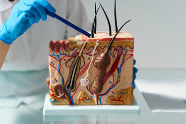
Human skin offers 1.5 to 2.0 m² of surface and contributes 15%–16% of body weight, which makes this organ the most reachable site for drug administration and the strictest gate in non-invasive therapeutics.1–3 Since scopolamine reached the market in patch form in 1979,4 the pharmacokinetic case has stayed persuasive: delivery bypasses hepatic first-pass metabolism, holds plasma concentrations steady, removes gastrointestinal degradation, and earns ready patient acceptance.5–7 Peptide and protein medicines, now more than one-tenth of the global pharmaceutical market, would gain most.8–10 Yet therapeutic macromolecules, which range from roughly 500 Da to beyond 150 kDa and are hydrophilic, charged at physiological pH, and vulnerable to proteolysis, sit opposite the small, moderately lipophilic, uncharged profile that passive permeation rewards.9–11 Virtually no therapeutic macromolecule meets those criteria.2,4,5
An Ordered Lipid Seal
The stratum corneum governs how rapidly and completely foreign molecules reach the circulation.2,4,12 Ten to 25 layers of anucleated corneocytes sit within a specialized lipid matrix, long pictured as bricks set in mortar.2,13 Permeability resides in the mortar, the sole continuous route open to an applied molecule.2,12,14 Ceramides, cholesterol, and free fatty acids occupy an approximately equimolar arrangement,12,14,15 while phospholipids are essentially absent, persisting only in trace amounts, a compositional oddity among mammalian membranes.4,7 Hydrocarbon chains adopt orthorhombic crystalline packing, the densest lateral state accessible to lipids and the least permeable;16,17 saturated fatty acids of C20–C24 chain length drive that arrangement, and shorter homologs fail to produce the same order. Ultra-long-chain acylceramides bridge adjacent bilayers and hold the lamellae together as molecular rivets.14
Geometry compounds chemistry. Because a permeant winds around successive corneocytes rather than crossing in a straight line, the effective diffusion distance reaches 450 to 500 μm despite a tissue thickness of 10 to 20 μm, a tortuosity factor of 25 to 50.18–20 Damage triggers immediate secretion of preformed lamellar-body lipids, which restores roughly half of barrier function within hours, while complete lamellar reorganization requires several days. That two-phase kinetic sets the clock on how long microchannels created by microneedles, electroporation, or laser ablation remain patent, typically 2 to 4 hours unoccluded and up to 72 hours under occlusion.4,21
Living Layers Add Resistance
Beneath the horny layer, the viable epidermis measures 50 to 100 μm across most body sites, and considerably thicker on the palm and sole;12,19 it holds roughly 40% protein and 40% water and constitutes a far more hydrophilic medium.22–24 Tight junctions built from claudin-1, claudin-4, claudin-7, occludin, and ZO-1 form a second gate within the granular layer, 25–27 regulating what fraction of an applied agent advances into deeper strata.26 Experimental work confirms that the full epidermis, not the horny layer alone, sets the ceiling on delivery.28 Capillary loops in the dermis reach within 100 to 200 μm of the surface29 and clear arriving molecules quickly, sustaining the inward gradient that drives absorption.30 Fine nociceptive fibers ascend into the epidermis, yet the dense dermal nerve plexus resides below approximately 1,000 μm; microneedle arrays shorter than 700 to 750 μm therefore stimulate few nociceptors and are reported as painless, which defines the practical insertion window.2,19,31
Takeaway 1: The barrier is a series of filters, not one membrane. Piercing the horny layer removes only the first, since tight junctions in the living epidermis continue to restrain water-soluble macromolecules.
Three Routes Of Permeation
The transcellular path runs through corneocytes but forces 10 to 20 sequential partitioning events between water-rich keratin and lipid domains,19,32,33 a sequence made hostile to peptides by lysine, arginine, aspartate, and glutamate residues.34 The intercellular route through the lipid mortar carries most permeants;2,12,19 extracting those lipids increases flux by several orders of magnitude and identifies the matrix as rate-limiting.7 Therapeutic peptides, with log P values commonly between −2 and −6, fall well outside the favorable window of 1 to 335,36 (Table 1). Aqueous microdomains within the matrix measure only 20 to 30 Å across,37 the paracellular gap in the living epidermis narrows to 3 to 10 Å,38 and partitioning between vehicle and horny layer lipids runs four to six orders of magnitude below that of ideal candidates5,8 (Figure 1). Hair follicles and sweat glands occupy a mere 0.1% of surface area,11,26,39 though electrical resistance through those structures falls 100- to 1,000-fold below that of the intact horny layer; ionic current therefore funnels preferentially through the appendages.26,39
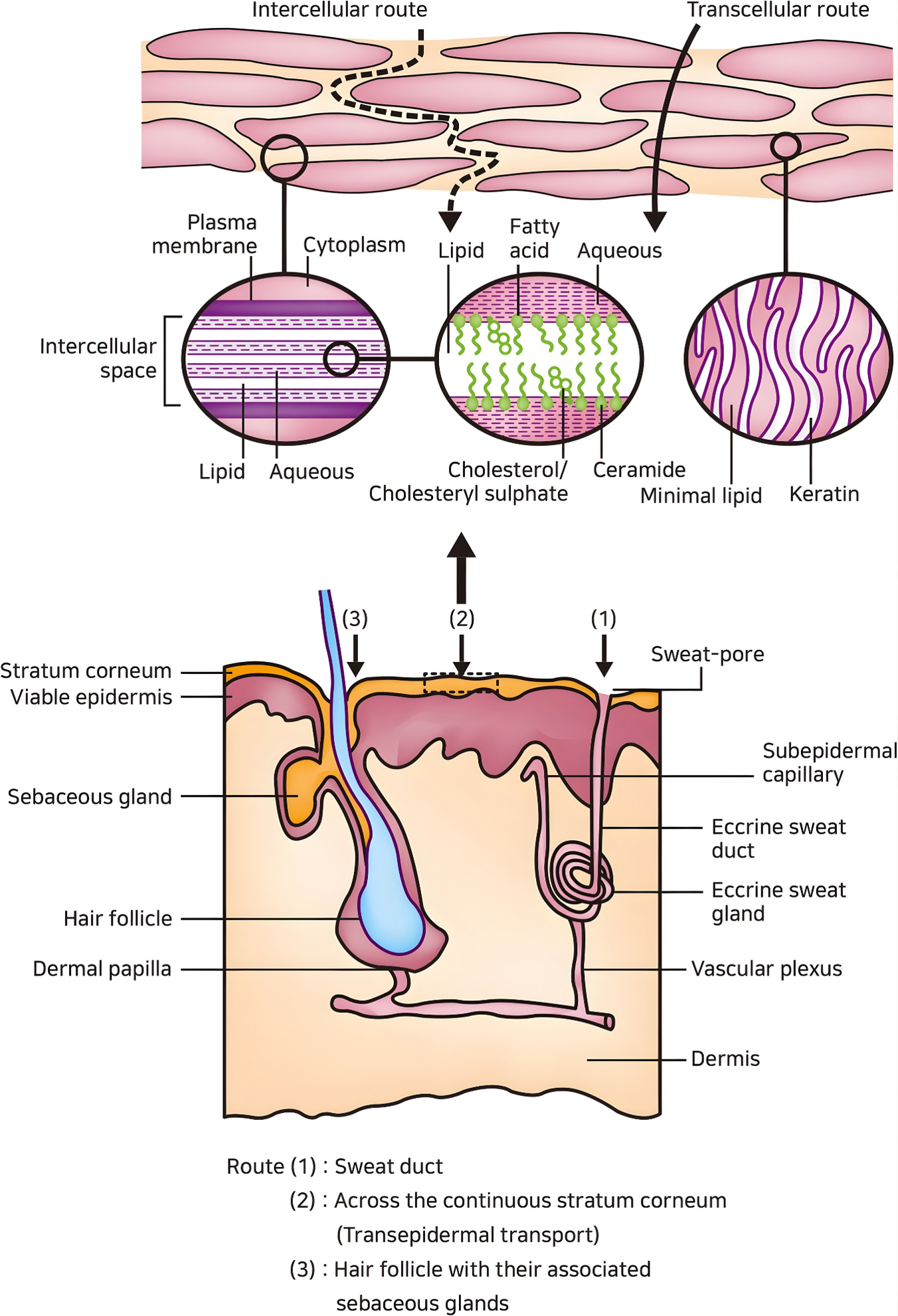
Figure 1: Organization of the horny layer and the three routes available to topically applied agents. Adapted with permission from Kim, B.; Cho, H.-E.; Moon, S. H.; Ahn, H.-J.; Bae, S.; Cho, H.-D.; An, S., Transdermal Delivery Systems in Cosmetics. biomed dermatol 2020, 4 (1), 10.40 [Creative Commons Attribution 4.0 International License.]
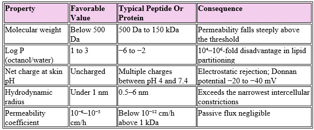
Table 1: Passive permeation requirements set against typical peptide and protein properties.
Predictive models capture the trend without resolving the peptide case. The Potts–Guy relationship correlates permeability with lipophilicity and mass,33,41 yet performs poorly for flexible peptides whose conformation and ionization shift with local conditions.5 Empirically, every additional 100 Da above 500 Da costs 50%–90% of permeability.42 Chemical modification helps only partially: palmitoylation lifted the log P of decapeptide-12 from −6.5 to −0.8, improving retention, though the modified peptide still fell short of the favorable window.43
Enzymes And Instability
Surviving the physical seal does not guarantee survival of the molecule. Kallikreins KLK5, KLK7, and KLK14 accept exogenous substrates and degrade part of an applied dose before inward diffusion proceeds,5,26 and the living epidermis holds a wider catalog, including cathepsins, aminopeptidases, carboxypeptidases, and esterases.2,5,7 Skin proteolytic activity runs roughly tenfold below intestinal activity, a real advantage over oral dosing, yet epidermal enzymes acting together still degrade insulin and comparable agents during permeation.7,44 Co-formulated protease inhibitors measurably increase delivery,45 while D-amino acid substitution, N-methylation, cyclization, PEGylation, and encapsulation within lipid nanocarriers reduce enzymatic attrition.46–48 Chemical instability adds deamidation at asparagine-glycine and asparagine-serine sequences49 and oxidation of exposed methionine, cysteine, and tryptophan,50 while aggregation follows nucleation kinetics in which slow oligomer formation precedes rapid fibril growth.51
Immunological Duality
Skin holds 800 to 1,200 antigen-presenting cells per mm², compared with roughly 200 per mm² in muscle. That density cuts two ways. Microneedle-mediated intradermal vaccination matches intramuscular immunogenicity at fourfold or greater dose reduction.4,8,52 Chronic replacement therapy faces the mirror image, since repeated intradermal dosing risks anti-drug antibodies that neutralize activity or provoke hypersensitivity.8,52,53 Aggregates behave as adjuvants and sharply increase immunogenicity relative to the native monomer,10,53 and proteins above 10 kDa carry the highest risk.54
Takeaway 2: The same cellular density that makes skin an excellent vaccination site makes skin a hazardous one for long-term protein replacement. Indication, not technology alone, should decide whether intradermal delivery suits a given molecule.
From Negligible Flux To Therapeutic Doses
Passive delivery yields 1%–5% bioavailability for small agents and essentially none above 1 kDa.5,26,41 Alternative noninvasive routes fare little better, with oral insulin reaching 0.81% and oral desmopressin 0.08–0.16%.9,41,55 Active enhancement changes the outcome decisively (Figure 2).
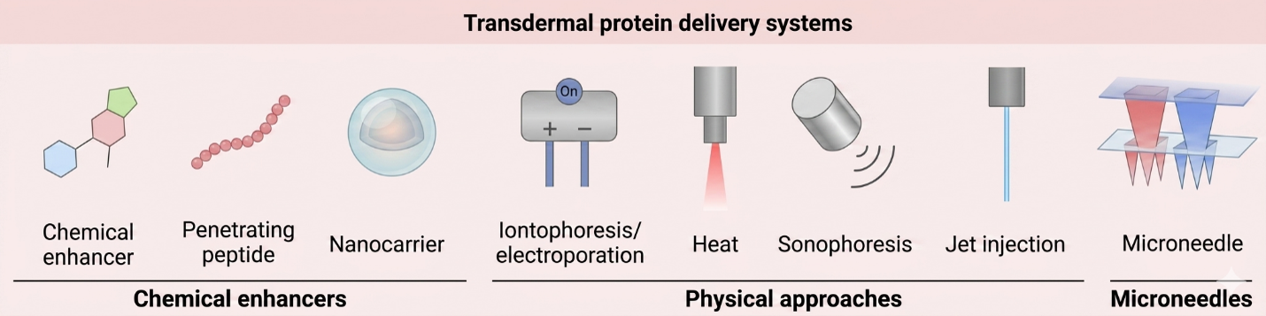
Figure 2: Active methods used to move protein therapeutics across skin, arranged by operating principle. Adapted with permission from Yang, Y.; Zhou, R.; Wang, Y.; Zhang, Y.; Yu, J.; Gu, Z., Recent Advances in Oral and Transdermal Protein Delivery Systems. Angewandte Chemie International Edition 2023, 62 (10), e202214795.55 Permission to reuse granted under license number 6363150371612.
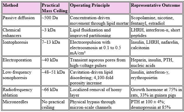
Table 2: Enhancement methods, molecular ceilings, and representative performance.
Combining methods yields more than the sum of the parts. Microneedle pretreatment followed by iontophoresis increased methotrexate delivery 25-fold and parathyroid hormone delivery 17-fold over iontophoresis alone, and electroporation preceding iontophoresis improved LHRH flux five- to tenfold.21,44 (Table 2). Practical ceilings persist, since surface tension and viscous resistance restrict loading to 1 to 5 μg per needle and about 1 mg per array.11,21 Economics narrow the field further, because low bioavailability multiplies dose and cost: oral insulin at 0.81% would demand roughly 120 times the injected dose, a burden untenable for recombinant proteins priced at $20 to $500 per milligram.41 High-potency agents dosed in micrograms, including parathyroid hormone, desmopressin, and vaccine antigens, therefore reached commercially credible pharmacokinetics first.8,21,45

Conclusion
The skin evolved to exclude water-soluble macromolecules and performs that task with high efficiency. Delivery science has answered not by defeating the barrier but by dismantling specific mechanisms: physical bypass of the lipid seal, electrokinetic driving forces through low-resistance channels, molecular redesign against proteases, and formulation chemistry against aggregation. Four priorities now deserve attention. Microneedle products require demonstrated bioequivalence to approved injectables and shelf stability free of cold chain dependence. Stimulus-responsive systems keyed to glucose, pH, or temperature would permit individualized dosing. Permeation models incorporating molecular dynamics and patient-specific barrier measurement would replace correlations that fail for flexible molecules. Finally, repeated barrier disruption calls for long-term safety data on immune homeostasis, microbial community structure, and infection risk.
References:
- Boer, M.; Duchnik, E.; Maleszka, R.; Marchlewicz, M. Structural and Biophysical Characteristics of Human Skin in Maintaining Proper Epidermal Barrier Function. Postepy Dermatol Alergol 2016, 33 (1), 1–5. https://doi.org/10.5114/pdia.2015.48037
- Jiménez-Rodríguez, A.; Guardado-Félix, D.; Antunes-Ricardo, M. Challenges and Strategies for Topical and Transdermal Delivery of Bioactive Peptides. Crit Rev Ther Drug Carrier Syst 2022, 39 (1), 1–31.
- Kolarsick, P. A. J.; Kolarsick, M. A.; Goodwin, C. Anatomy and Physiology of the Skin. Journal of the Dermatology Nurses’ Association 2011, 3 (4), 203–213. https://doi.org/10.1097/JDN.0b013e3182274a98
- Nguyen, H. X. Microneedles: The Future of Drug Delivery; CRC Press, 2023.
- Bhavsar, J.; Kasture, K.; Salvi, B. V.; Shende, P. Strategies for Transportation of Peptides across the Skin for Treatment of Multiple Diseases. Ther Deliv 2025, 16 (1), 63–86. https://doi.org/10.1080/20415990.2024.2411943
- Peña-Juárez, M.; Guadarrama-Escobar, O. R.; Escobar-Chávez, J. J. Transdermal Delivery Systems for Biomolecules. Journal of Pharmaceutical Innovation 2021, 1–14.
- Shah, B.; Surti, N.; Misra, A. Other Routes of Protein and Peptide Delivery: Transdermal, Topical, Uterine, and Rectal. In Challenges in Delivery of Therapeutic Genomics and Proteomics; Elsevier, 2011; pp 623–671. https://doi.org/10.1016/B978-0-12-384964-9.00012-8
- Liu, T.; Chen, M.; Fu, J.; Sun, Y.; Lu, C.; Quan, G.; Pan, X.; Wu, C. Recent Advances in Microneedles-Mediated Transdermal Delivery of Protein and Peptide Drugs. Acta Pharmaceutica Sinica B 2021, 11 (8), 2326–2343.
- Wang, L.; Wang, N.; Zhang, W.; Cheng, X.; Yan, Z.; Shao, G.; Wang, X.; Wang, R.; Fu, C. Therapeutic Peptides: Current Applications and Future Directions. Signal transduction and targeted therapy 2022, 7 (1), 48.
- Wu, J.; Sahoo, J. K.; Li, Y.; Xu, Q.; Kaplan, D. L. Challenges in Delivering Therapeutic Peptides and Proteins: A Silk-Based Solution. Journal of Controlled Release 2022, 345, 176–189.
- Long, L.; Zhang, J.; Yang, Z.; Guo, Y.; Hu, X.; Wang, Y. Transdermal Delivery of Peptide and Protein Drugs: Strategies, Advantages and Disadvantages. Journal of Drug Delivery Science and Technology 2020, 60, 102007. https://doi.org/10.1016/j.jddst.2020.102007
- Bouwstra, J. A.; Nădăban, A.; Bras, W.; McCabe, C.; Bunge, A.; Gooris, G. S. The Skin Barrier: An Extraordinary Interface with an Exceptional Lipid Organization. Prog Lipid Res 2023, 92, 101252. https://doi.org/10.1016/j.plipres.2023.101252
- Cheng, T.; Tai, Z.; Shen, M.; Li, Y.; Yu, J.; Wang, J.; Zhu, Q.; Chen, Z. Advance and Challenges in the Treatment of Skin Diseases with the Transdermal Drug Delivery System. Pharmaceutics 2023, 15 (8), 2165. https://doi.org/10.3390/pharmaceutics15082165
- Berdyshev, E. Skin Lipid Barrier: Structure, Function and Metabolism. Allergy Asthma Immunol Res 2024, 16 (5), 445–461. https://doi.org/10.4168/aair.2024.16.5.445
- Ohno, Y.; Nakamura, T.; Iwasaki, T.; Katsuyama, A.; Ichikawa, S.; Kihara, A. Determining the Structure of Protein-Bound Ceramides, Essential Lipids for Skin Barrier Function. iScience 2023, 26 (11), 108248. https://doi.org/10.1016/j.isci.2023.108248
- Bouwstra, J. A.; Gooris, G. S. The Lipid Organisation in Human Stratum Corneum and Model Systems. 2010.
- Radhakrishnan, N.; Kaul, S. C.; Wadhwa, R.; Yang, L.-W.; Sundar, D. Establishing a General Atomistic Model for the Stratum Corneum Lipid Matrix Based on Experimental Data for Skin Permeation Studies. Int J Mol Sci 2025, 26 (2), 674. https://doi.org/10.3390/ijms26020674
- Bouwstra, J. A.; Honeywell-Nguyen, P. L.; Gooris, G. S.; Ponec, M. Structure of the Skin Barrier and Its Modulation by Vesicular Formulations. Prog Lipid Res 2003, 42 (1), 1–36. https://doi.org/10.1016/s0163-7827(02)00028-0
- Liu, M.; Chen, S.; Zhang, Z.; Li, H.; Sun, G.; Yin, N.; Wen, J. Anti-Ageing Peptides and Proteins for Topical Applications: A Review. Pharm Dev Technol 2022, 27 (1), 108–125. https://doi.org/10.1080/10837450.2021.2023569
- Zaid Alkilani, A.; McCrudden, M. T.; Donnelly, R. F. Transdermal Drug Delivery: Innovative Pharmaceutical Developments Based on Disruption of the Barrier Properties of the Stratum Corneum. Pharmaceutics 2015, 7 (4), 438–470.
- Kalluri, H.; Banga, A. K. Transdermal Delivery of Proteins. AAPS PharmSciTech 2011, 12 (1), 431–441. https://doi.org/10.1208/s12249-011-9601-6
- Alexander, A.; Dwivedi, S.; Giri, T. K.; Saraf, S.; Saraf, S.; Tripathi, D. K. Approaches for Breaking the Barriers of Drug Permeation through Transdermal Drug Delivery. J. Control. Release. 2012, 164 (1), 26–40. https://doi.org/10.1016/j.jconrel.2012.09.017
- Jaitley, S.; Saraswathi, T. Pathophysiology of Langerhans Cells. J Oral Maxillofac Pathol 2012, 16 (2), 239–244. https://doi.org/10.4103/0973-029X.99077
- Matsui, T.; Amagai, M. Dissecting the Formation, Structure and Barrier Function of the Stratum Corneum. Int Immunol 2015, 27 (6), 269–280. https://doi.org/10.1093/intimm/dxv013
- Chen, J.; Liu, C.; Yang, Y.; Gong, X.; Qian, H. The Stratum Corneum Barrier: Impaired Function in Relation to Associated Lipids and Proteins. Tissue Barriers 2025, 13 (2), 2361197. https://doi.org/10.1080/21688370.2024.2361197
- Drug Delivery in Dermatology: Fundamental and Practical Applications; Kalil, C. L. P. V., Campos, V., Eds.; Springer International Publishing: Cham, 2021. https://doi.org/10.1007/978-3-030-81807-4
- Nguyen, A. V.; Soulika, A. M. The Dynamics of the Skin’s Immune System. Int J Mol Sci 2019, 20 (8), 1811. https://doi.org/10.3390/ijms20081811
- Andrews, S. N.; Jeong, E.; Prausnitz, M. R. Transdermal Delivery of Molecules Is Limited by Full Epidermis, Not Just Stratum Corneum. Pharmaceutical research 2013, 30 (4), 1099–1109.
- Dj, T. Biochemistry of Human Skin--Our Brain on the Outside. Chemical Society reviews 2006, 35 (1), 52–67. https://doi.org/10.1039/b505793k
- Korosec, A.; Frech, S.; Gesslbauer, B.; Vierhapper, M.; Radtke, C.; Petzelbauer, P.; Lichtenberger, B. M. Lineage Identity and Location within the Dermis Determine the Function of Papillary and Reticular Fibroblasts in Human Skin. J Invest Dermatol 2019, 139 (2), 342–351. https://doi.org/10.1016/j.jid.2018.07.033
- Aich, K.; Singh, T.; Dang, S. Advances in Microneedle-Based Transdermal Delivery for Drugs and Peptides. Drug Delivery and Translational Research 2022, 12 (7), 1556–1568.
- Barbero, A. M.; Frasch, H. F. Transcellular Route of Diffusion through Stratum Corneum: Results from Finite Element Models. J Pharm Sci 2006, 95 (10), 2186–2194. https://doi.org/10.1002/jps.20695
- Thakur, N.; Thakur, S.; Singh, Y. P.; Prasad, S.; Kaur, B.; Sharma, C. A Current Review on Transdermal Delivery of Protein and Peptides: Possibilities and Challenges. Letters in Drug Design & Discovery 2024, 21 (19), 4597–4605. https://doi.org/10.2174/0115701808357267250102054810
- Bos, J. D.; Meinardi, M. M. The 500 Dalton Rule for the Skin Penetration of Chemical Compounds and Drugs. Experimental Dermatology 2000, 9 (3), 165–169.
- Muheem, A.; Shakeel, F.; Jahangir, M. A.; Anwar, M.; Mallick, N.; Jain, G. K.; Warsi, M. H.; Ahmad, F. J. A Review on the Strategies for Oral Delivery of Proteins and Peptides and Their Clinical Perspectives. Saudi Pharm J 2016, 24 (4), 413–428. https://doi.org/10.1016/j.jsps.2014.06.004
- Verma, S.; Goand, U. K.; Husain, A.; Katekar, R. A.; Garg, R.; Gayen, J. R. Challenges of Peptide and Protein Drug Delivery by Oral Route: Current Strategies to Improve the Bioavailability. Drug Dev Res 2021, 82 (7), 927–944. https://doi.org/10.1002/ddr.21832
- Mitragotri, S.; Anissimov, Y. G.; Bunge, A. L.; Frasch, H. F.; Guy, R. H.; Hadgraft, J.; Kasting, G. B.; Lane, M. E.; Roberts, M. S. Mathematical Models of Skin Permeability: An Overview. Int J Pharm 2011, 418 (1), 115–129. https://doi.org/10.1016/j.ijpharm.2011.02.023
- Bajracharya, R.; Song, J. G.; Back, S. Y.; Han, H.-K. Recent Advancements in Non-Invasive Formulations for Protein Drug Delivery. Computational and structural biotechnology journal 2019, 17, 1290–1308.
- Hasan, M.; Khatun, A.; Kogure, K. Iontophoresis of Biological Macromolecular Drugs. Pharmaceutics 2022, 14 (3), 525. https://doi.org/10.3390/pharmaceutics14030525
- Kim, B.; Cho, H.-E.; Moon, S. H.; Ahn, H.-J.; Bae, S.; Cho, H.-D.; An, S. Transdermal Delivery Systems in Cosmetics. biomed dermatol 2020, 4 (1), 10. https://doi.org/10.1186/s41702-020-0058-7
- Brown, L. R. Commercial Challenges of Protein Drug Delivery. Expert Opin Drug Deliv 2005, 2 (1), 29–42. https://doi.org/10.1517/17425247.2.1.29
- Potts, R. O.; Guy, R. H. A Predictive Algorithm for Skin Permeability: The Effects of Molecular Size and Hydrogen Bond Activity. Pharmaceutical research 1995, 12 (11), 1628–1633.
- Chen, J.; Bian, J.; Hantash, B. M.; Albakr, L.; Hibbs, D. E.; Xiang, X.; Xie, P.; Wu, C.; Kang, L. Enhanced Skin Retention and Permeation of a Novel Peptide via Structural Modification, Chemical Enhancement, and Microneedles. Int J Pharm 2021, 606, 120868. https://doi.org/10.1016/j.ijpharm.2021.120868
- Benson, H. A. E.; Namjoshi, S. Proteins and Peptides: Strategies for Delivery to and across the Skin. J Pharm Sci 2008, 97 (9), 3591–3610. https://doi.org/10.1002/jps.21277
- Herwadkar, A.; Banga, A. K. Peptide and Protein Transdermal Drug Delivery. Drug Discovery Today: Technologies 2012, 9 (2), e147–e154. https://doi.org/10.1016/j.ddtec.2011.11.007
- Dragulska, S. A.; Chen, Y.; Wlodarczyk, M. T.; Poursharifi, M.; Dottino, P.; Ulijn, R. V.; Martignetti, J. A.; Mieszawska, A. J. Tripeptide-Stabilized Oil-in-Water Nanoemulsion of an Oleic Acids-Platinum(II) Conjugate as an Anticancer Nanomedicine. Bioconjug Chem 2018, 29 (8), 2514–2519. https://doi.org/10.1021/acs.bioconjchem.8b00409
- Jirwankar, P.; Shah, D.; Shao, J. Protection of Protein Drugs by Self-Emulsified Nanoemulsion Against Proteolysis. J Pharm Sci 2020, 109 (8), 2615–2621. https://doi.org/10.1016/j.xphs.2020.05.005
- Yamada, M.; Tayeb, H.; Wang, H.; Dang, N.; Mohammed, Y. H.; Osseiran, S.; Belt, P. J.; Roberts, M. S.; Evans, C. L.; Sainsbury, F.; Prow, T. W. Using Elongated Microparticles to Enhance Tailorable Nanoemulsion Delivery in Excised Human Skin and Volunteers. J Control Release 2018, 288, 264–276. https://doi.org/10.1016/j.jconrel.2018.09.012
- Robinson, N. E.; Robinson, A. B. Molecular Clocks. Proc Natl Acad Sci U S A 2001, 98 (3), 944–949. https://doi.org/10.1073/pnas.98.3.944
- Swain, S.; Mondal, D.; Beg, S.; Patra, C. N.; Dinda, S. C.; Sruti, J.; Rao, M. E. B. Stabilization and Delivery Approaches for Protein and Peptide Pharmaceuticals: An Extensive Review of Patents. Recent Pat Biotechnol 2013, 7 (1), 28–46. https://doi.org/10.2174/1872208311307010004
- Roberts, C. J. Therapeutic Protein Aggregation: Mechanisms, Design, and Control. Trends Biotechnol 2014, 32 (7), 372–380. https://doi.org/10.1016/j.tibtech.2014.05.005
- Priyanka, M.; Reddy, M. S.; Vijetha, K. A. Transdermal Patches for Peptides, Vaccines and Biologics: Current Progress and Challenges. 2025, 03 (08). https://doi.org/10.5281/zenodo.16851658
- Tan, M. L.; Choong, P. F.; Dass, C. R. Recent Developments in Liposomes, Microparticles and Nanoparticles for Protein and Peptide Drug Delivery. Peptides 2010, 31 (1), 184–193.
- Crommelin, D. J. A.; Storm, G.; Verrijk, R.; de Leede, L.; Jiskoot, W.; Hennink, W. E. Shifting Paradigms: Biopharmaceuticals versus Low Molecular Weight Drugs. Int J Pharm 2003, 266 (1–2), 3–16. https://doi.org/10.1016/s0378-5173(03)00376-4
- Yang, Y.; Zhou, R.; Wang, Y.; Zhang, Y.; Yu, J.; Gu, Z. Recent Advances in Oral and Transdermal Protein Delivery Systems. Angewandte Chemie International Edition 2023, 62 (10), e202214795. https://doi.org/10.1002/anie.202214795
About The Author:
 Hiep X. Nguyen, DPharm, Ph.D., DTM, is a faculty member in Pharmaceutical & Biomedical Sciences at California Northstate University. With over a decade developing topical, transdermal, and injectable products, he holds a Ph.D. from Mercer University and a pharmacy degree from Hanoi University of Pharmacy. A Distinguished Toastmaster and certified pharmacist, he serves as co-director of the Vietnamese Association of Pharmacists & Pharmaceutical Scientists, USA, and advises PharmD and PhD students. Nguyen has authored 30+ peer-reviewed articles, a seminal book (Microneedles: The Future of Drug Delivery), and five book chapters. Recognized among Stanford University’s top 2% of scientists globally, he has delivered numerous international presentations and over 34 scientific abstracts.