
Microneedles In Drug Delivery: From Bench To Patch
By Hiep X. Nguyen, DPharm, Ph.D., DTM, California Northstate University
The pharmaceutical sector continues to pursue delivery methods that combine patient convenience with therapeutic efficacy. Parenteral products dominate new drug approvals but suffer from needle phobia affecting roughly 20% of the population, sharps disposal challenges, and cold-chain dependencies.1,2 Oral administration, while preferred for its convenience, struggles with enzymatic degradation and poor absorption.3 Transdermal delivery offers a promising middle ground, yet the stratum corneum — a 10–20 µm outermost layer of corneocytes embedded within a lipid-rich matrix — restricts permeation to moderately lipophilic compounds with log P values between 1.0 and 3.0.4,5
Microneedle (MN) technology has emerged as a third-generation transdermal system that overcomes this skin barrier through micron-scale projections, typically 25 to 2000 µm in length, that create channels into the viable epidermis without reaching pain-sensitive nerve endings or blood vessels. Despite its potential, no microneedle array patch product for therapeutic drug delivery has received marketing authorization from the FDA or EMA to date.6 This article presents the scientific principles, technological progress, and translational hurdles defining the present state of microneedle research.
Classification of Microneedle Systems
Six categories define the MN field, each differing in payload localization, release kinetics, and delivery mechanism:
- Solid microneedles create transient hydrophilic channels via a “poke and patch” method, after which a drug formulation diffuses passively through the micropores.7 Pore closure kinetics — sensitive to formulation characteristics and drug physicochemistry — define the delivery window.8
- Coated microneedles carry a thin dry film of active pharmaceutical ingredient on solid needle surfaces and release the payload within minutes of skin insertion.9,10
- Hollow microneedles function as miniaturized hypodermic needles, enabling controlled infusion or interstitial fluid (ISF) extraction through an open lumen.11,12
- Dissolving microneedles, fabricated from water-soluble polymers or biodegradable matrices, release encapsulated agents upon hydration by ISF, eliminating sharps waste.13,14
- Hydrogel-forming swelling microneedles use crosslinked polymer networks that imbibe ISF and form diffusive conduits between drug reservoirs and dermal capillaries.15,16
- Porous microneedles feature interconnected micro- and nanoscale channels that accommodate substantial drug payloads.6,17
Below, Table 1 describes comparative features of microneedle types.
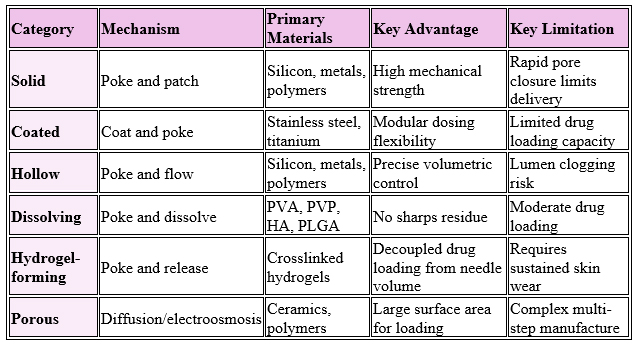
Table 1: Comparative features of microneedle types.
Practical takeaway: Match the MN type to the therapeutic goal: dissolving MN for vaccines and biologics, hydrogel-forming MN for sustained delivery, and hollow MN for diagnostics or bolus dosing.
Skin Properties Guiding Microneedle Design
Human skin presents a nonlinear, anisotropic, heterogeneous, and viscoelastic mechanical barrier that any microneedle must overcome reliably.18 The stratum corneum has a shear modulus near 20 MPa, the epidermis a Young’s modulus between 1 and 10 MPa, and the dermis varies in thickness from 0.9 to 1.7 mm across body sites.19,20 Collagen fiber families within the dermis align along Langer’s lines, generating direction-dependent stiffness whereby modulus along these lines reaches 112.5 MPa, roughly 176% of perpendicular values.21,22 Viscoelasticity carries practical implications for MN insertion. Failure stress increases from 12 MPa at a strain rate of 0.06 s⁻¹ to 19 MPa at 167 s⁻¹, indicating that dynamic loading demands higher peak forces but exploits rate-dependent stiffening to achieve consistent puncture.23
MN geometry directly governs insertion mechanics. Smaller tip radii concentrate stress and lower the puncture threshold; flat-tipped nickel needles with diameters from 30 to 80 µm showed insertion forces scaling linearly from 0.08 to 3.04 N with tip area.24 However, tips that are too sharp risk plastic deformation, particularly in polymer needles.25 MN body morphology shapes both penetration efficiency and structural durability, with wall thickness in hollow needles raising fracture force from 0.5 to 5.5 N when increased from 5 to 58 µm.24,26 Inter-needle spacing produces the “bed-of-nails” effect: closely packed arrays distribute load and reduce per-needle penetration depth.27
Practical takeaway: Design needles with tip diameters below 15 µm, body lengths between 200 and 900 µm, and inter-needle spacing of at least 150 µm to balance penetration efficiency with manufacturing feasibility.
Materials And Manufacturing Methods
Material selection balances biocompatibility, mechanical performance, and processability.
- Silicon offered the original substrate due to lithographic accessibility, but remains brittle and now serves mostly for master molds.28,29
- Metals such as stainless steel, titanium, and nickel deliver superior toughness, though may generate harmful tip residues.30
- Polymers dominate present research, with poly(lactic-co-glycolic acid), chitosan, and hyaluronic acid leading the field due to safety, biodegradability, and tunable dissolution kinetics.31–33
- Hyaluronic acid, a natural polysaccharide widely used in cosmetic MN products, offers tunable mechanical properties based on molecular weight.34,35
- Silk fibroin enables both mechanically strong tips and dissolvable needles.36,37
Fabrication methods divide into four categories: molding, additive manufacturing, etching, and forming.
- Micromolding remains the dominant industrial method, with vacuum, compression, and centrifugal methods accommodating polymer melts and solutions.38–40
- Additive technologies, i.e., stereolithography (SLA), digital light processing (DLP), two-photon polymerization (TPP), and continuous liquid interface production (CLIP), now produce tip diameters below 5 µm with print times ranging from minutes to hours.41–43
- Etching achieves the sharpest features but demands cleanroom infrastructure.44,45
- Forming methods, including hot embossing and magnetorheological drawing, offer minute-scale cycle times suitable for scale-up.46,47
Below, Table 2 describes representative fabrication methods and scalability characteristics.
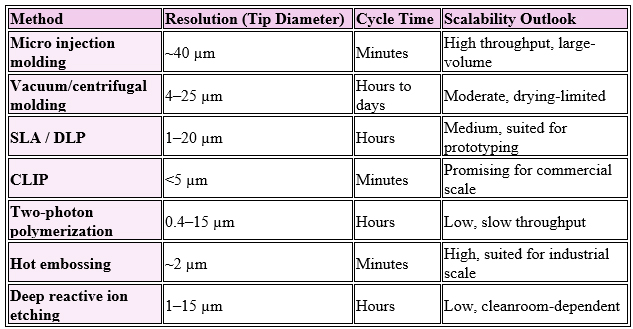
Table 2: Representative fabrication methods and scalability characteristics.
Practical takeaway: Prioritize hot embossing or CLIP for high-throughput commercial scale-up; reserve etching and two-photon methods for prototyping and validation studies.
Innovative Designs And Stimulus-Responsive Systems
Microneedle research has progressed beyond passive delivery toward dynamic, programmable systems. Stimulus-responsive systems release drugs upon exposure to pH, light, temperature, magnetic fields, electric stimuli, mechanical force, glucose, or enzymes.48–50 Glucose-responsive MN patches use either glucose oxidase catalysis or phenylboronic acid recognition to release insulin on demand during hyperglycemia, offering closed-loop diabetes management.51–53
Iontophoretic microneedles combine physical penetration with electric repulsion and electroosmotic flow to enhance the delivery of hydrophilic and macromolecular drugs.54,55 Bionic designs draw inspiration from mosquito mouthparts, bee stings, parasitoid wasps, and snake teeth to minimize insertion force and refine retention. Examples include shark-tooth-inspired wound patches and dual-engine bionic microneedles that improved bioavailability and neurological function in Alzheimer’s models.56,57 Core–shell microneedles use a mechanically robust outer shell encasing a drug-laden core, decoupling mechanical performance from drug-release kinetics.
Applications
Microneedles offer promising applications in vaccination, sustained drug delivery, biologics administration, biosensing, and cosmetic therapy. The Vaccine Innovation Prioritization Strategy, a partnership among Gavi, WHO, the Gates Foundation, UNICEF, and PATH, has identified MN patches as a priority innovation due to dose-sparing immunogenicity and reduced cold-chain demand.58 Clinical trials of influenza, measles, and rubella MN vaccines have demonstrated favorable safety and immunogenicity profiles.59–61
Long-acting delivery has progressed substantially. Yavuz and colleagues achieved 100-day in vitro release of levonorgestrel from silk fibroin needles, while microparticle incorporation extended release beyond one year.62 Effervescent microneedles enabled rapid backing separation and sustained delivery exceeding one month in rats.63 HIV antiretrovirals (rilpivirine, cabotegravir) have reached therapeutic plasma levels for up to 56 days following patch removal.64,65
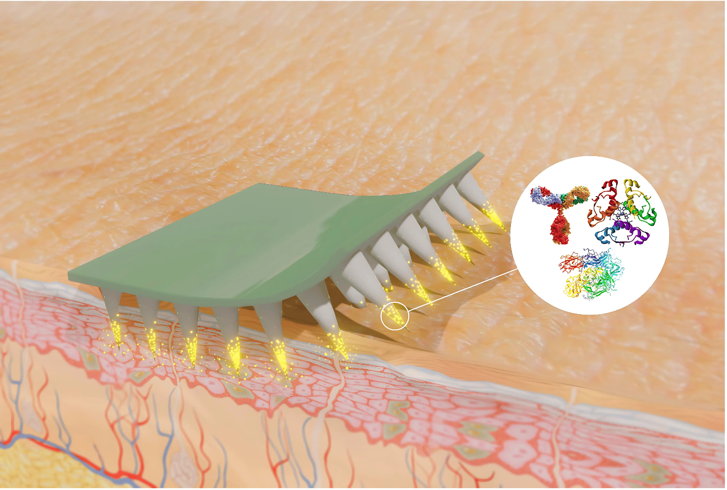
Figure 1: Microneedle application in transdermal delivery of macromolecules. Image reprinted with permission from reference 66, under the Creative Commons (CC) BY license.
For biologics, microneedles enable the delivery of antibodies, peptides, hormones, and nucleic acids in dry-state formulations that bypass first-pass metabolism (Figure 1 above).66,67 The Soluvia hollow MN system facilitated intradermal influenza vaccination (Fluzone Intradermal, Intanza), while the SCS Microinjector (Xipere) delivers triamcinolone acetonide to the suprachoroidal space for uveitic macular edema68 (refer to Table 3 below).
Diagnostic applications have reached commercial milestones. Biolinq Shine received FDA De Novo classification in September 2025 as the first fully autonomous needle-free continuous glucose monitor. YourBio Health’s TAP blood-collection devices secured 510(k) clearances for both professional and layperson use. Electrochemical MN sensors now monitor multiple metabolites simultaneously, while wristband-integrated systems combine ISF chemistry with ECG and ultrasonic cardiovascular signals.69,70.
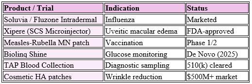
Table 3: Selected clinical and commercial milestones.
Practical takeaway: Vaccination, long-acting contraception, and continuous biosensing represent the highest-value near-term opportunities for MN commercialization.

Applicator Systems
Reliable insertion across diverse skin types and anatomical sites demands more than careful needle design. Quasi-static applicators rely on user-applied force, while impact-based devices store elastic energy in springs, deformable structures, or electromagnetic actuators released by mechanical triggers.71,72 Assistive methods such as vibration and skin pre-stretch lower the energy required for penetration by concentrating stress at needle tips.73–75 Effective applicators must work across age groups, body mass indices, and anatomical sites while providing tactile, auditory, or visual confirmation of successful insertion.76,77
Translational Challenges
Despite substantial preclinical progress, several barriers slow commercial translation:
- Drug loading capacity remains limited for small-array patches, particularly for coated MN, where excess coating compromises mechanical performance.78
- Sterilization presents acute challenges, as dry heat, steam, and gamma irradiation may damage thermolabile biologics; viable options include ethylene oxide treatment, low-dose gamma radiation or electron-beam irradiation (for radiation-tolerant payloads), or aseptic processing.79
- Regulatory pathways add further complexity. MN combination products require concurrent evaluation of device performance and drug stability, often extending review timelines by roughly 30% relative to standalone devices.80 Standalone diagnostic MNs may follow 510(k) or De Novo pathways, while drug-loaded patches fall under combination product oversight.81
- Toxicity assessments have indicated the biological safety of various materials. Carbohydrate-based polymers, including carboxymethyl cellulose, maltodextrin, and hyaluronic acid, showed negligible cytotoxicity across concentrations from 3 to 80 mg/mL.82 Skin irritation, when reported, correlates with needle length, material composition, and drug payload.83
Practical takeaway: Engage regulators early through pre-IND or Q-Submission meetings, and adopt quality by design principles to streamline combination product review.
Future Outlook
The next decade will likely witness the first marketing authorization of an MN drug delivery product, driven by maturing manufacturing infrastructure, refined regulatory science, and growing clinical evidence. Computational modeling and machine learning are accelerating design optimization, with decision tree regression, Bayesian techniques, and neural networks predicting fluid collection efficiency, drug permeation, and skin penetration.84,85 Integration with wearable electronics and closed-loop dosing systems will enable theranostic systems that sense biomarkers and dispense therapeutics in real time.86 Self-administration capability stands at the center of this trajectory, particularly for low- and middle-income countries where contraceptive, antiretroviral, and vaccine MN products could materially expand access.76,77
Conclusion
Microneedle technology occupies a unique position at the intersection of materials science, biomechanics, pharmaceutical formulation, and medical device engineering. The field has matured from silicon prototypes to a diverse family of solid, coated, hollow, dissolving, hydrogel-forming, porous, and stimulus-responsive designs, each tailored to specific therapeutic and diagnostic objectives. Scalable manufacturing, predictive computational design, and harmonized regulatory pathways now define the critical path forward. With clinical evidence accumulating and the first commercial diagnostic MN devices already reaching the market, microneedles stand poised to broaden their role across vaccination, long-acting drug delivery, biologics administration, and personalized healthcare. For drug delivery leaders, the strategic imperative is clear: invest in scalable manufacturing methods, partner early with regulators, and align development programs with high-value clinical indications where MN technology delivers definitive advantages over conventional dosage forms.
References
- Donnelly RF, Prausnitz MR. The promise of microneedle technologies for drug delivery. Drug Deliv Transl Res. 2024;14(3):573-580. doi:10.1007/s13346-023-01430-8
- Simoens S, Huys I. R&D Costs of New Medicines: A Landscape Analysis. Front Med (Lausanne). 2021;8:760762. doi:10.3389/fmed.2021.760762
- Pawar K. Recent Updates in Vaccine Delivery through Microneedles. Adv Pharm Bull. 2023;13(1):1-4. doi:10.34172/apb.2023.001
- Roberts MS, Cheruvu HS, Mangion SE, et al. Topical drug delivery: History, percutaneous absorption, and product development. Advanced Drug Delivery Reviews. 2021;177:113929. doi:10.1016/j.addr.2021.113929
- Sabbagh F, Kim BS. Recent advances in polymeric transdermal drug delivery systems. J Control Release. 2022;341:132-146. doi:10.1016/j.jconrel.2021.11.025
- Zhang J, Li H, Albakr L, et al. Microneedle-enabled therapeutics delivery and biosensing in clinical trials. J Control Release. 2023;360:687-704. doi:10.1016/j.jconrel.2023.07.023
- Li WZ, Huo MR, Zhou JP, et al. Super-short solid silicon microneedles for transdermal drug delivery applications. Int J Pharm. 2010;389(1-2):122-129. doi:10.1016/j.ijpharm.2010.01.024
- Ogunjimi AT, Lawson C, Carr J, Patel KK, Ferguson N, Brogden NK. Micropore Closure Rates following Microneedle Application at Various Anatomical Sites in Healthy Human Subjects. Skin Pharmacol Physiol. 2021;34(4):214-228. doi:10.1159/000515454
- Chen X, Kask AS, Crichton ML, et al. Improved DNA vaccination by skin-targeted delivery using dry-coated densely-packed microprojection arrays. Journal of controlled release. 2010;148(3):327-333.
- Zhang Y, Brown K, Siebenaler K, Determan A, Dohmeier D, Hansen K. Development of lidocaine-coated microneedle product for rapid, safe, and prolonged local analgesic action. Pharm Res. 2012;29(1):170-177. doi:10.1007/s11095-011-0524-4
- Gade S, Glover K, Mishra D, et al. Hollow microneedles for ocular drug delivery. J Control Release. 2024;371:43-66. doi:10.1016/j.jconrel.2024.05.013
- Jiang X, Jin Y, Zeng Y, Shi P, Li W. Self-Implantable Core-Shell Microneedle Patch for Long-Acting Treatment of Keratitis via Programmed Drug Release. Small. 2024;20(29):e2310461. doi:10.1002/smll.202310461
- Ali M, Namjoshi S, Benson HA, Mohammed Y, Kumeria T. Dissolvable polymer microneedles for drug delivery and diagnostics. Journal of Controlled Release. 2022;347:561-589.
- Yang ZR, Suo H, Fan JW, et al. Endogenous stimuli-responsive separating microneedles to inhibit hypertrophic scar through remodeling the pathological microenvironment. Nat Commun. 2024;15(1):2038. doi:10.1038/s41467-024-46328-2
- Donnelly RF, McCrudden MT, Alkilani AZ, et al. Hydrogel-forming microneedles prepared from “super swelling” polymers combined with lyophilised wafers for transdermal drug delivery. PloS one. 2014;9(10):e111547.
- Turner JG, White LR, Estrela P, Leese HS. Hydrogel-Forming Microneedles: Current Advancements and Future Trends. Macromolecular Bioscience. 2021;21(2):2000307. doi:10.1002/mabi.202000307
- Bao L, Park J, Bonfante G, Kim B. Recent advances in porous microneedles: materials, fabrication, and transdermal applications. Drug Deliv Transl Res. 2022;12(2):395-414. doi:10.1007/s13346-021-01045-x
- Wei JC, Edwards GA, Martin DJ, Huang H, Crichton ML, Kendall MA. Allometric scaling of skin thickness, elasticity, viscoelasticity to mass for micro-medical device translation: from mice, rats, rabbits, pigs to humans. Scientific reports. 2017;7(1):1-16.
- Feng X, Li GY, Ramier A, Eltony AM, Yun SH. In vivo stiffness measurement of epidermis, dermis, and hypodermis using broadband Rayleigh-wave optical coherence elastography. Acta Biomater. 2022;146:295-305. doi:10.1016/j.actbio.2022.04.030
- Shu W, Heimark H, Bertollo N, Tobin DJ, O’Cearbhaill ED, Annaidh AN. Insights into the mechanics of solid conical microneedle array insertion into skin using the finite element method. Acta Biomater. 2021;135:403-413. doi:10.1016/j.actbio.2021.08.045
- Ní Annaidh A, Bruyère K, Destrade M, Gilchrist MD, Otténio M. Characterization of the anisotropic mechanical properties of excised human skin. J Mech Behav Biomed Mater. 2012;5(1):139-148. doi:10.1016/j.jmbbm.2011.08.016
- Rosicka K, Hill M, Wdowski MM. Skin anisotropy: Finding the optimal incision line for volar forearm in males and females. J Mech Behav Biomed Mater. 2021;124:104805. doi:10.1016/j.jmbbm.2021.104805
- Ottenio M, Tran D, Ní Annaidh A, Gilchrist MD, Bruyère K. Strain rate and anisotropy effects on the tensile failure characteristics of human skin. J Mech Behav Biomed Mater. 2015;41:241-250. doi:10.1016/j.jmbbm.2014.10.006
- Davis SP, Landis BJ, Adams ZH, Allen MG, Prausnitz MR. Insertion of microneedles into skin: measurement and prediction of insertion force and needle fracture force. J Biomech. 2004;37(8):1155-1163.
- Takehara H, Inada M, Kanda Y, Ichiki T. Mechanical finite element analysis of needle tip shape to develop insertable polymer-based microneedle without plastic deformation. J Mech Behav Biomed Mater. 2025;163:106885. doi:10.1016/j.jmbbm.2025.106885
- Bolton CJW, Howells O, Blayney GJ, et al. Hollow silicon microneedle fabrication using advanced plasma etch technologies for applications in transdermal drug delivery. Lab Chip. 2020;20(15):2788-2795. doi:10.1039/d0lc00567c
- Jahan S, Jain A, Fregonese S, Hu C, Bacca M, Panat R. Bed-of-Nails effect: Unraveling the insertion behavior of aerosol jet 3D printed microneedle array in soft tissue. Extreme Mechanics Letters. 2025;77:102301. doi:10.1016/j.eml.2025.102301
- Chang H, Zheng M, Chew SWT, Xu C. Advances in the formulations of microneedles for manifold biomedical applications. Advanced Materials Technologies. 2020;5(4):1900552.
- Faraji Rad Z, Prewett PD, Davies GJ. An overview of microneedle applications, materials, and fabrication methods. Beilstein J Nanotechnol. 2021;12:1034-1046. doi:10.3762/bjnano.12.77
- Zhang R, Miao Q, Deng D, Wu J, Miao Y, Li Y. Research progress of advanced microneedle drug delivery system and its application in biomedicine. Colloids and Surfaces B: Biointerfaces. 2023;226:113302. doi:10.1016/j.colsurfb.2023.113302
- Jung JH, Jin SG. Microneedle for transdermal drug delivery: current trends and fabrication. Journal of Pharmaceutical Investigation. 2021;51(5):503-517.
- Nguyen HX, Kipping T, Banga AK. Polymeric Microneedles Enhance Transdermal Delivery of Therapeutics. Pharmaceutics. 2024;16(7):845.
- Saha I, Rai VK. Hyaluronic acid based microneedle array: Recent applications in drug delivery and cosmetology. Carbohydrate Polymers. 2021;267:118168. doi:10.1016/j.carbpol.2021.118168
- Jang M, Baek S, Kang G, Yang H, Kim S, Jung H. Dissolving microneedle with high molecular weight hyaluronic acid to improve skin wrinkles, dermal density and elasticity. Int J Cosmet Sci. 2020;42(3):302-309. doi:10.1111/ics.12617
- Kang H, Zuo Z, Lin R, Yao M, Han Y, Han J. The most promising microneedle device: present and future of hyaluronic acid microneedle patch. Drug Deliv. 2022;29(1):3087-3110. doi:10.1080/10717544.2022.2125600
- Tsioris K, Raja WK, Pritchard EM, Panilaitis B, Kaplan DL, Omenetto FG. Fabrication of Silk Microneedles for Controlled-Release Drug Delivery. Advanced functional materials. 2012;22(2):330-335.
- Zhu M, Liu Y, Jiang F, Cao J, Kundu SC, Lu S. Combined Silk Fibroin Microneedles for Insulin Delivery. ACS Biomater Sci Eng. 2020;6(6):3422-3429. doi:10.1021/acsbiomaterials.0c00273
- 3Lee HS, Ryu HR, Roh JY, Park JH. Bleomycin-coated microneedles for treatment of warts. Pharmaceutical research. 2017;34(1):101-112.
- Nejad HR, Sadeqi A, Kiaee G, Sonkusale S. Low-cost and cleanroom-free fabrication of microneedles. Microsyst Nanoeng. 2018;4(1):17073. doi:10.1038/micronano.2017.73
- Yin Y, Su W, Zhang J, et al. Separable microneedle patch to protect and deliver DNA nanovaccines against COVID-19. ACS nano. 2021;15(9):14347-14359.
- Faraji Rad Z, Prewett PD, Davies GJ. Parametric optimization of two-photon direct laser writing process for manufacturing polymeric microneedles. Additive Manufacturing. 2022;56:102953. doi:10.1016/j.addma.2022.102953
- Johnson AR, Caudill CL, Tumbleston JR, et al. Single-Step Fabrication of Computationally Designed Microneedles by Continuous Liquid Interface Production. PLoS One. 2016;11(9):e0162518. doi:10.1371/journal.pone.0162518
- Ovsianikov A, Chichkov B, Mente P, Monteiro-Riviere NA, Doraiswamy A, Narayan RJ. Two Photon Polymerization of Polymer–Ceramic Hybrid Materials for Transdermal Drug Delivery. International Journal of Applied Ceramic Technology. 2007;4(1):22-29. doi:10.1111/j.1744-7402.2007.02115.x
- Pradeep Narayanan S, Raghavan S. Fabrication and characterization of gold-coated solid silicon microneedles with improved biocompatibility. Int J Adv Manuf Technol. 2019;104(9):3327-3333. doi:10.1007/s00170-018-2596-3
- Tabassum N, Alba M, Yan L, Voelcker NH. Porous Silicon Microneedles for Enhanced Transdermal Drug Delivery. Advanced Therapeutics. Published online 2022:2200156.
- Andersen TE, Andersen AJ, Petersen RS, Nielsen LH, Keller SS. Drug loaded biodegradable polymer microneedles fabricated by hot embossing. Microelectronic Engineering. 2018;195:57-61. doi:10.1016/j.mee.2018.03.024
- Chen Z, Ren L, Li J, et al. Rapid fabrication of microneedles using magnetorheological drawing lithography. Acta Biomater. 2018;65:283-291. doi:10.1016/j.actbio.2017.10.030
- Chen X, Yu H, Wang L, Shen D, Li C, Zhou W. Cross-Linking-Density-Changeable Microneedle Patch Prepared from a Glucose-Responsive Hydrogel for Insulin Delivery. ACS Biomater Sci Eng. 2021;7(10):4870-4882. doi:10.1021/acsbiomaterials.1c01073
- Gowda BHJ, Ahmed MG, Sahebkar A, Riadi Y, Shukla R, Kesharwani P. Stimuli-Responsive Microneedles as a Transdermal Drug Delivery System: A Demand-Supply Strategy. Biomacromolecules. 2022;23(4):1519-1544. doi:10.1021/acs.biomac.1c01691
- Shan J, Zhang X, Kong B, et al. Coordination polymer nanozymes-integrated colorimetric microneedle patches for intelligent wound infection management. Chemical Engineering Journal. 2022;444:136640. doi:10.1016/j.cej.2022.136640
- Chen S, Miyazaki T, Itoh M, et al. A Porous Reservoir-Backed Boronate Gel Microneedle for Efficient Skin Penetration and Sustained Glucose-Responsive Insulin Delivery. Gels. 2022;8(2):74. doi:10.3390/gels8020074
- Chen W, Tian R, Xu C, et al. Microneedle-array patches loaded with dual mineralized protein/peptide particles for type 2 diabetes therapy. Nature communications. 2017;8(1):1-11.
- Yu J, Wang J, Zhang Y, et al. Glucose-responsive insulin patch for the regulation of blood glucose in mice and minipigs. Nat Biomed Eng. 2020;4(5):499-506. doi:10.1038/s41551-019-0508-y
- Abe H, Sato K, Kimura N, et al. Porous Microneedle Patch for Electroosmosis-Promoted Transdermal Delivery of Drugs and Vaccines. Advanced NanoBiomed Research. 2022;2(1):2100066. doi:10.1002/anbr.202100066
- Luo X, Yu Q, Liu Y, et al. Closed-Loop Diabetes Minipatch Based on a Biosensor and an Electroosmotic Pump on Hollow Biodegradable Microneedles. ACS Sens. 2022;7(5):1347-1360. doi:10.1021/acssensors.1c02337
- Raikar AS, Kalaskar DM, Bhilegaonkar S, Somnache SN, Bodaghi M. Revolutionizing drug delivery by bioinspired 4D transdermal microneedles: Advances and future horizons. European Polymer Journal. 2024;210:112952. doi:10.1016/j.eurpolymj.2024.112952
- Zhang M, Yang B, Ren T, et al. Dual engine-driven bionic microneedles for early intervention and prolonged treatment of Alzheimer’s disease. Journal of Controlled Release. 2024;367:184-196. doi:10.1016/j.jconrel.2024.01.030
- Feng YX, Hu H, Wong YY, Yao X, He ML. Microneedles: An Emerging Vaccine Delivery Tool and a Prospective Solution to the Challenges of SARS-CoV-2 Mass Vaccination. Pharmaceutics. 2023;15(5):5. doi:10.3390/pharmaceutics15051349
- Adigweme I, Akpalu E, Yisa M, et al. Study protocol for a phase 1/2, single-centre, double-blind, double-dummy, randomized, active-controlled, age de-escalation trial to assess the safety, tolerability and immunogenicity of a measles and rubella vaccine delivered by a microneedle patch in healthy adults (18 to 40 years), measles and rubella vaccine-primed toddlers (15 to 18 months) and measles and rubella vaccine-naïve infants (9 to 10 months) in The Gambia [Measles and Rubella Vaccine Microneedle Patch Phase 1/2 Age De-escalation Trial]. Trials. 2022;23(1):775. doi:10.1186/s13063-022-06493-5
- Adigweme I, Yisa M, Ooko M, et al. A measles and rubella vaccine microneedle patch in The Gambia: a phase 1/2, double-blind, double-dummy, randomised, active-controlled, age de-escalation trial. Lancet. 2024;403(10439):1879-1892. doi:10.1016/S0140-6736(24)00532-4
- Forster AH, Witham K, Depelsenaire ACI, et al. Safety, tolerability, and immunogenicity of influenza vaccination with a high-density microarray patch: Results from a randomized, controlled phase I clinical trial. PLoS Medicine. 2020;17(3):e1003024. doi:10.1371/journal.pmed.1003024
- Yavuz B, Chambre L, Harrington K, Kluge J, Valenti L, Kaplan DL. Silk Fibroin Microneedle Patches for the Sustained Release of Levonorgestrel. ACS Appl Bio Mater. 2020;3(8):5375-5382. doi:10.1021/acsabm.0c00671
- Li W, Tang J, Terry RN, et al. Long-acting reversible contraception by effervescent microneedle patch. Science advances. 2019;5(11):eaaw8145.
- Moffatt K, Tekko IA, Vora L, et al. Development and Evaluation of Dissolving Microarray Patches for Co-administered and Repeated Intradermal Delivery of Long-acting Rilpivirine and Cabotegravir Nanosuspensions for Paediatric HIV Antiretroviral Therapy. Pharm Res. 2023;40(7):1673-1696. doi:10.1007/s11095-022-03408-6
- Volpe-Zanutto F, Vora LK, Tekko IA, et al. Hydrogel-forming microarray patches with cyclodextrin drug reservoirs for long-acting delivery of poorly soluble cabotegravir sodium for HIV Pre-Exposure Prophylaxis. J Control Release. 2022;348:771-785. doi:10.1016/j.jconrel.2022.06.028
- Nguyen HX, Nguyen CN. Microneedle-Mediated Transdermal Delivery of Biopharmaceuticals. Pharmaceutics. 2023;15(1):1. doi:10.3390/pharmaceutics15010277
- Kim E, Erdos G, Huang S, et al. Microneedle array delivered recombinant coronavirus vaccines: Immunogenicity and rapid translational development. EBioMedicine. 2020;55:102743.
- Cárcamo-Martínez Á, Mallon B, Domínguez-Robles J, Vora LK, Anjani QK, Donnelly RF. Hollow microneedles: A perspective in biomedical applications. International Journal of Pharmaceutics. 2021;599:120455. doi:10.1016/j.ijpharm.2021.120455
- Chang AY, Lin M, Yin L, et al. Integration of chemical and physical inputs for monitoring metabolites and cardiac signals in diabetes. Nat Biomed Eng. 2026;10(1):94-109. doi:10.1038/s41551-025-01439-z
- Tehrani F, Teymourian H, Wuerstle B, et al. An integrated wearable microneedle array for the continuous monitoring of multiple biomarkers in interstitial fluid. Nat Biomed Eng. 2022;6(11):1214-1224. doi:10.1038/s41551-022-00887-1
- Kang G, Kim M, Yang H, et al. Latch Applicator for Efficient Delivery of Dissolving Microneedles Based on Rapid Release of Elastic Strain Energy by Thumb Force. Advanced Functional Materials. 2023;33(11):2210805. doi:10.1002/adfm.202210805
- Leone M, Van Oorschot BH, Nejadnik MR, et al. Universal Applicator for Digitally-Controlled Pressing Force and Impact Velocity Insertion of Microneedles into Skin. Pharmaceutics. 2018;10(4):4. doi:10.3390/pharmaceutics10040211
- Ebrahiminejad V, Malek-khatabi A, Faraji Rad Z. Influence of Low-Frequency Vibration and Skin Strain on Insertion Mechanics and Drug Diffusion of PVA/PVP Dissolving Microneedles. Advanced Materials Technologies. 2024;9(4):2301272. doi:10.1002/admt.202301272
- Kim J, Park S, Nam G, Choi Y, Woo S, Yoon SH. Bioinspired microneedle insertion for deep and precise skin penetration with low force: Why the application of mechanophysical stimuli should be considered. J Mech Behav Biomed Mater. 2018;78:480-490. doi:10.1016/j.jmbbm.2017.12.006
- Kim M, Kang G, Min HS, Lee Y, Park S, Jung H. Evolution of microneedle applicators for vaccination: the role of the latch applicator in optimizing dissolving microneedle-based immunization. Expert Opin Drug Deliv. 2024;21(12):1823-1835. doi:10.1080/17425247.2024.2422939
- El-Sahn M, Elliott R, El-Sahn M, Lucas J, Wood Santos T. End-user research into understanding perceptions of and reactions to a microarray patch (MAP) for contraception among women in Ghana, Kenya and Uganda. Front Reprod Health. 2024;6:1351692. doi:10.3389/frph.2024.1351692
- Gualeni B, Hughes L, Stauber I, et al. Human-centred design of a new microneedle-based hormonal contraceptive delivery system. Gates Open Res. 2021;5:96. doi:10.12688/gatesopenres.13233.3
- Ai X, Yang J, Liu Z, Guo T, Feng N. Recent progress of microneedles in transdermal immunotherapy: A review. International Journal of Pharmaceutics. 2024;662:124481. doi:10.1016/j.ijpharm.2024.124481
- Dul M, Alali M, Ameri M, et al. Assessing the risk of a clinically significant infection from a Microneedle Array Patch (MAP) product. J Control Release. 2023;361:236-245. doi:10.1016/j.jconrel.2023.07.001
- Sartawi Z, Blackshields C, Faisal W. Dissolving microneedles: Applications and growing therapeutic potential. Journal of Controlled Release. 2022;348:186-205. doi:10.1016/j.jconrel.2022.05.045
- Avcil M, Çelik A. Microneedles in Drug Delivery: Progress and Challenges. Micromachines (Basel). 2021;12(11):1321. doi:10.3390/mi12111321
- Yalcintas EP, Ackerman DS, Korkmaz E, et al. Analysis of In Vitro Cytotoxicity of Carbohydrate-Based Materials Used for Dissolvable Microneedle Arrays. Pharm Res. 2020;37(3):33. doi:10.1007/s11095-019-2748-7
- Ghosh TK. Dermal Drug Delivery: From Innovation to Production. CRC Press; 2020.
- Biswas AA, Dhondale MR, Agrawal AK, Serrano DR, Mishra B, Kumar D. Advancements in microneedle fabrication techniques: artificial intelligence assisted 3D-printing technology. Drug Deliv and Transl Res. 2024;14(6):1458-1479. doi:10.1007/s13346-023-01510-9
- Tarar C, Aydın E, K. Yetisen A, Tasoglu S. Bayesian machine learning optimization of microneedle design for biological fluid sampling. Sensors & Diagnostics. 2023;2(4):858-866. doi:10.1039/D3SD00103B
- Moonla C, Reynoso M, Chang AY, Saha T, Surace S, Wang J. Microneedle-Based Multiplexed Monitoring of Diabetes Biomarkers: Capabilities Beyond Glucose Toward Closed-Loop Theranostic Systems. ACS Sens. 2025;10(8):5363-5379. doi:10.1021/acssensors.5c00652
About The Author:
 Hiep X. Nguyen, DPharm, Ph.D., DTM, is a faculty member in Pharmaceutical & Biomedical Sciences at California Northstate University. With over a decade developing topical, transdermal, and injectable products, he holds a Ph.D. from Mercer University and a pharmacy degree from Hanoi University of Pharmacy. A Distinguished Toastmaster and certified pharmacist, he serves as co-director of the Vietnamese Association of Pharmacists & Pharmaceutical Scientists, USA, and advises PharmD and PhD students. Nguyen has authored 30+ peer-reviewed articles, a seminal book (Microneedles: The Future of Drug Delivery), and five book chapters. Recognized among Stanford University’s top 2% of scientists globally, he has delivered numerous international presentations and over 34 scientific abstracts.